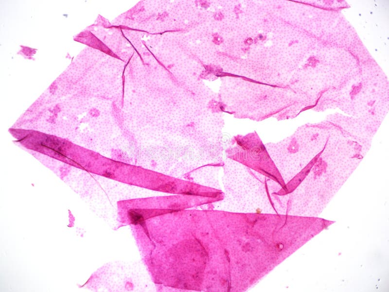
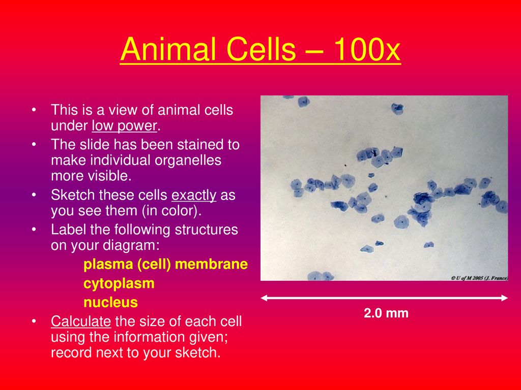
animal cell under microscope 100x
Pond water refers to a standing body of water. The micrographs in Figures 2 and 3 were obtained using 100X 600X 1000x and 1500X pHase Contrast Dark.
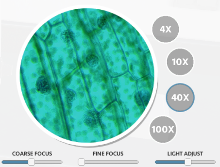
Typical Animal Cell Center 40x Stock Image Image Of Typical Science 152965947
A nucleus or a cell wall can be seen more clearly by using different stains.

. Together these data suggest that malignant cells from MBM express a neuronal-like phenotype which may be important for brain-metastatic. Also overheating causes damage to the structure of the cell which would affect the end results. Diatom Neo-Latin diatoma refers to any member of a large group comprising several genera of algae specifically microalgae found in the oceans waterways and soils of the worldLiving diatoms make up a significant portion of the Earths biomass.
These include 3 multi-investigator groups that operate principally in the TBHIV space 2 extramural research units of the South African Medical Research Council and a number of other research entities 6 Research Chairs awarded under the National Research Foundations South African Research Chairs Initiative SARChI and further areas of. Brightfield Light Microscope Compound light microscope This is the most basic optical Microscope used in microbiology laboratories which produces a dark image against a bright background. In addition the electron microscope is required to resolve the structure of mitochondria bacteria viruses and large protein complexes.
At 100x magnification you will be able to see 2mm. These are characteristics of living cells that are capable of division and growth. At 400x magnification you will be able to see 045mm or 450 microns.
Single-cell segmentation and quantification revealed a significantly higher fraction of cancer cells expressing NCAM1 and higher NCAM1 intensity in melanoma cells from MBMs Figures 3O 3P and S4HS4K. Each polyp is a sac-like. Pond Water Under the Microscope.
To see the cell organelles you will need to get a higher magnification usually with a 40x-100x objective lens. SPM was founded in 1981 with the invention of the scanning tunneling microscope an instrument for imaging surfaces at the atomic levelThe first successful scanning tunneling microscope experiment was done by Gerd Binnig and. While some can be seen with the naked eye others are too small and will require the use of a microscope to be able to properly observe.
Condensers are lenses that are used to collect and focus light from the illuminator into the specimen. Took up SR101 and dynamically spread it across the tumor cell network while GBCs lacking anatomical connections barely took up SR101 Figures 1B and 1CThese mutually connected GBCs showed multicellular co-active calcium events indicating functional connectivity across this GBC subpopulation while calcium events remained local in GBCs not exhibiting connecting. At 1000x magnification you will be able to see 0180mm or 180 microns.
At 40x magnification you will be able to see 5mm. Bottom left and right under a 100 optical microscope and the average number of cells was calculated. Unlike epidermal cells of various plants epidermal cells of.
When you are ready challenge your knowledge in the testing section to see what you have learned. Silicon chloride and calcium identified under an ESEM microscope coupled with an EDS x-ray microprobe. The specimen is most often an ultrathin section less than 100 nm thick or a suspension on a grid.
Under the microscope animal cells appear different based on the type of the cell. P21 loss partially rescued EdU incorporation Figure 4N while having no effect on Arg replete cells demonstrating that a GCN2-mediated p21 induction contributes to HCC cell-cycle arrest. Under a higher magnification of 100X nuclei of the cells appear.
Transmission electron microscopy TEM is a microscopy technique in which a beam of electrons is transmitted through a specimen to form an image. They are found under the stage next to the diaphragm of the microscope. Animal cell under the microscope.
The human hepatoellular carcinomas cell line HepG2 Hep3B and Huh7 were obtained from ACTT with ACTTnumber. To assess the contribution of p21 to cell-cycle arrest we silenced p21 using shRNAs capable of lowering mRNA and protein levels 80 Figures 4L and 4M. When viewed under the microscope Gram negative and Gram positive bacteria will produce different results.
Differentiate between a condenser and an Abbe condenser. The images of Paulownia wood hair and frogs blood were captured with a high power compound microscope using a Nikon camera adapter. Pond water contains a variety of plant and animal life.
Cell Wall Structure and Gram Stain. This research has focused largely on CD8 T cells with a focus on both those antigens that are recognised and the means by which they are presented. Iodine crystal violet and methylene blue are examples of simple stains.
A typical animal cell is 1020 μm in diameter which is about one-fifth the size of the smallest particle visible to the naked eye. When viewed under the microscope it is possible to view the cell nucleus a very thin layer of cytoplasm that can be seen in some of the cells as well as the cell walls at the boundary of each cell. X axis KeV Y axis Counts7583.
Lesson Description BioNetworks Virtual Microscope is the first fully interactive 3D scope - its a great practice tool to prepare you for working in a science lab. His research has centered on understanding the mechanisms by which the human immune system recognises the Mycobacterium tuberculosis Mtb infected cell. Microscope cell staining is a technique used to improve the visibility of cells and cell parts under a microscope.
Fluorescence Microscope - study the most used microscope in medicalbiological fields which uses high powered light waves to provide unique image viewing options. Explore topics on usage care terminology and then interact with a fully functional virtual microscope. Corals are marine invertebrates within the class Anthozoa of the phylum CnidariaThey typically form compact colonies of many identical individual polypsCoral species include the important reef builders that inhabit tropical oceans and secrete calcium carbonate to form a hard skeleton.
Here the authors perform a genome-wide CRISPR-Cas9 knockout screen to systematically identify and characterize essential and growth-restricting genes in human trophoblast cells. Dark Field Microscope - learn more about how when the light source is blocked off light scatters as it hits the specimen and is then able to reveal details otherwise difficult to see. A coral group is a colony of very many genetically identical polyps.
They generate about 20 to 50 percent of the oxygen produced on the planet each year take in over 67 billion metric tons of. The compound microscope typically has three or four magnifications - 40x 100x 400x and sometimes 1000x. Scanning probe microscopy SPM is a branch of microscopy that forms images of surfaces using a physical probe that scans the specimen.
Using fetal organ cells and animal organ cell lines undisclosed kept hidden as trade. Made up of two lenses it is widely used to view plant and animal cell organelles including some parasites such as Paramecium after staining with basic stains. Make a wet or dry mount with a coverslip.
Inhibition rate of the drug on cell migration 1. They play a major role in ensuring clear sharp images are produced with a high magnification of 400X and above. At 100x magnification you will be able to see 2mm.
HB-8065 HB-8064 and PTA-4583. Cell lines and cell culture conditions. An image is formed from the interaction of the electrons with the sample as the beam is transmitted through the specimen.
You can see a variety of cells pretty well with the light microscope. This work is licensed under a Creative Commons Attribution-NonCommercial-NoDerivs 25 LicenseCreative Commons Attribution-NonCommercial-NoDerivs 25 License. As well excessive decolorization can end up removing the primary stain.
This is usually smaller than a lake and may either be man-made or natural.

Microscopic View Of Algal Cells Cultured With And Without Co 2 Under Download Scientific Diagram
Virtual Microscope

Stem Of Cotton Center 100x Magnification Stock Image Image Of Cells Cytoplasm 152965833

180 100x Magnification Stock Photos Pictures Royalty Free Images Istock

Low 100x And High 400x Magnification Images Of Hematoxylin And Download Scientific Diagram

Histological Sections Of Zebrafish Ovaries Under 100x Magnification Of Download Scientific Diagram

Root Bacteria Side 100x Magnification Stock Image Image Of Biology Side 152966295
Virtual Microscope Animal And Plant Cell Tutorial Ppt Download
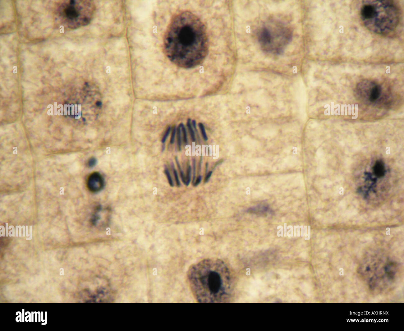
Mitosis Anaphase In Onion Tissue At 1000x Under Optical Microscope Inmersion Oil Objetive 100x Stock Photo Alamy

Blood Smear Show Platelet Increaseplatelet More Than 25 Cells Per 100x Microscope Stock Photo Download Image Now Istock
Typical Animal Cell Center 400x Stock Image Image Of Visible Compound 152965979
Starfish Embryology Fertilized Animal Egg Cell Shows Nucleus Cell Nucleus Starfish
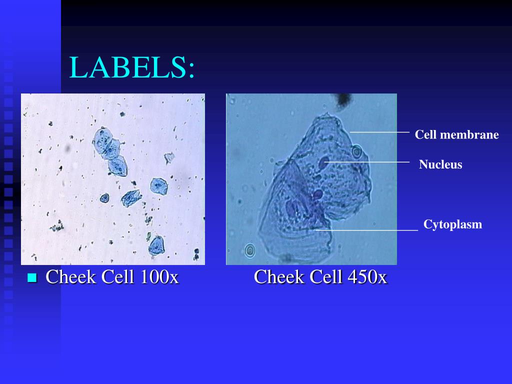
Ppt Post Lab Plant Animal Cells Or Powerpoint Presentation Free Download Id 5665034

Microscopy Micrograph Animal Conjugation Of Paramecium Caudatum Magnification 100x Stock Photo Picture And Royalty Free Image Image 16195330
![]()
Typical Plant Cell 100x Magnification Stock Image Image Of School College 152965951

180 100x Magnification Stock Photos Pictures Royalty Free Images Istock
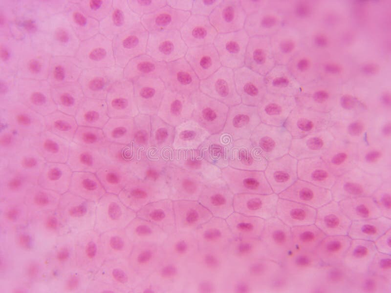
Typical Animal Cell Center 100x Stock Photo Image Of 100x School 152965862
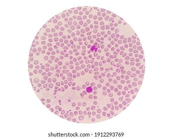
Lemur Blood Smear Under 100x Light Stock Photo 1912293769 Shutterstock
![]()
Typical Plant Cell 100x Magnification Stock Image Image Of Cells Magnification 152965909